GLP-1 Triggers a Brain-Gut-Heart Circuit That Could Prevent Post-Heart Attack ‘No-Reflow’
Up to half of patients whose blocked coronary artery is reopened still suffer poor blood flow to the heart muscle. Early research now points to a surprising three-way circuit connecting the gut, the brain, and the heart's tiniest blood vessels as a possible fix.

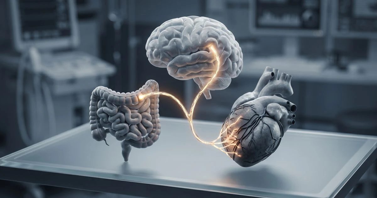
Up to 50% of patients who receive treatment to unblock a coronary artery still experience a frustrating phenomenon called microvascular 'no-reflow': the large artery opens, but blood fails to reach the heart muscle through the fine capillary network beneath it. The result is larger infarct size, lower left ventricular ejection fraction, and higher mortality, even after technically successful intervention. A new review published in June 2026 argues that the incretin hormone GLP-1 may address this problem through a mechanism far more intricate than anyone had expected, one that runs through the gut, the vagus nerve, and a class of capillary-wall cells called pericytes.
GLP-1's KATP Channel Action on Coronary Pericytes
The central mechanistic claim is that GLP-1 protects ischemic heart tissue by activating ATP-sensitive potassium channels (KATP channels) specifically on coronary pericytes. Pericytes are small contractile cells that wrap around capillaries and regulate their diameter. When pericytes constrict in response to ischemia-reperfusion injury, they can physically close off capillaries even after the upstream artery has been cleared, producing the no-reflow state. According to findings from the underlying experimental work, opening pericyte KATP channels with GLP-1 causes coronary capillary dilation, and blocking or genetically deleting those channels abolishes the effect entirely, directly implicating the channel as the effector, not merely a bystander.
This places pericytes, long studied in the brain and retina but historically underappreciated in the coronary circulation, at the center of a cardioprotective story.
The Brain-Gut-Heart Circuit That Drives Capillary Dilation
What makes this framework genuinely novel is not just the pericyte angle but the upstream signaling chain the authors describe. When skeletal muscle becomes ischemic, it sends signals that trigger GLP-1 release from intestinal L-cells in the gut. That gut-derived GLP-1 then travels via the vagus nerve to influence cardiac capillary tone, creating what the paper calls a brain-gut-heart pathway. Vagally-mediated GLP-1 release, in other words, appears capable of dilating coronary capillaries through the same pericyte KATP channel mechanism that pharmacological GLP-1 receptor agonists exploit.
This framing matters because it reframes GLP-1 not just as a diabetes or weight-loss drug but as a physiological signal that the body itself deploys during cardiovascular stress. The experiments used male C57BL mice, including knockout models in which pericyte KATP channels were genetically deleted, to confirm that the capillary dilation was channel-dependent rather than a broader hemodynamic effect.
Why This Matters for the Half of Patients Who Still Suffer After Angioplasty
The clinical stakes are substantial. Coronary microvascular obstruction after reperfusion is not a rare edge case: it affects roughly half of all myocardial infarction patients who undergo artery-opening procedures, and it independently predicts adverse outcomes regardless of how well the epicardial artery is treated. Existing evidence on GLP-1 receptor agonists in cardiovascular disease already shows that these drugs reduce risk across conditions including atherosclerosis, heart failure, and stroke, well beyond their original indication for type 2 diabetes. But the mechanisms behind those clinical benefits have remained incompletely understood.
If the pericyte-KATP channel pathway holds up in further testing, it would offer a specific, targetable mechanism for preventing no-reflow: drugs that selectively open pericyte KATP channels in the coronary microcirculation could, in principle, be administered at the time of reperfusion to keep capillaries patent. The review also catalogs a broader roster of GLP-1 receptor agonist actions in the myocardium, including suppression of proinflammatory cytokines such as IL-1β, TNF-α, IL-6, and MCP-1; inhibition of multiple cardiomyocyte death pathways (apoptosis, necroptosis, pyroptosis, and ferroptosis); and augmentation of autophagy and mitophagy to clear damaged cellular components.
Open Questions About GLP-1 Receptor Expression and Indirect Actions
The research is preliminary, and significant questions remain. A core unresolved issue in the field concerns where GLP-1 receptors are actually expressed in the heart. As one review of GLP-1 cardiovascular mechanisms has noted, GLP1R expression appears at low levels in the heart and vasculature, raising the question of whether GLP-1 acts directly on cardiac tissue or whether many of its benefits are mediated indirectly through neural, hormonal, or gut-derived intermediaries. The pericyte KATP channel story is itself partly an indirect mechanism: it depends on vagal signaling from the gut rather than GLP-1 acting on the cardiomyocyte itself.
Separately, a large meta-analysis in the European Heart Journal found significant reductions in major adverse cardiovascular events with GLP-1 receptor agonists but flagged substantial heterogeneity across trials and called for deeper investigation into mechanistic pathways that operate independently of glycemic control. The current paper's mouse-model data cannot resolve those translational questions. The studies used only male mice, leaving open how the pathway functions in female animals or in humans with the kinds of metabolic and vascular comorbidities typical of real heart attack patients.
What Comes Next in Pericyte-Targeted Cardioprotection
The authors suggest that pharmacological therapies targeting pericyte KATP channels could become a meaningful strategy for improving post-infarct recovery, particularly for the large proportion of patients who experience no-reflow despite successful reperfusion. The logical next steps would include replication in larger animal models, confirmation that the vagally-mediated gut-brain-heart signaling circuit operates in humans under ischemic conditions, and eventually trials of pericyte-targeted or GLP-1-based interventions delivered at the time of reperfusion rather than chronically.
For now, the paper reframes pericytes as active participants in coronary microvascular regulation rather than passive bystanders, and it positions the gut-vagus axis as a physiological cardioprotective system that pharmaceutical GLP-1 receptor agonists may be co-opting. That is a conceptual shift worth watching, even if the clinical application remains years away.
Evidence rating
INVESTIGATIONAL — 71/100
Caveats:
- All experiments were conducted in male C57BL mice, including knockout models — no human data exists yet.
- This is early-stage animal research; the brain–gut–heart pathway described has not been tested in clinical trials.
- Funding source was not disclosed by the authors.
Preclinical study conducted in male C57BL mice, including genetic knockout models. Demonstrates a mechanistic causal chain — vagally-mediated gut GLP-1 release activating pericyte KATP channels to induce coronary capillary dilation — but all findings are animal-only and no human data are presented. The pathway is novel and not yet independently replicated in humans.