Blocking One Receptor Fully Protected Aging Gut Stem Cells in Mice
A Nature Aging mouse study traces the decline of intestinal stem cells to systemic inflammation: TNF acting on TNFR1 in the gut lining switches off fat-burning genes and cuts the crypts' metabolic reserve. Young mice lacking that one receptor were completely protected from an aged circulation.

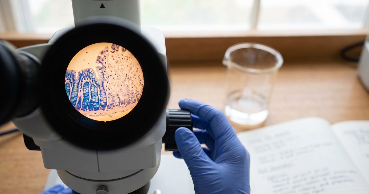
If your digestion has become less reliable with age, or you are managing a bowel condition and want to know what drives the decline, a new mouse study points somewhere unexpected: not the gut itself, but the inflammatory tone of the whole body. Researchers at Albert Einstein College of Medicine traced the age-related failure of intestinal stem cells to a single inflammatory messenger arriving from outside the intestine, and showed that deleting one receptor on the gut lining protected those stem cells completely. The work is entirely in mice, but it reframes gut aging as a systemic problem with a specific, druggable address.
Key Takeaways
- Intestinal crypts from aged mice produced about 30% fewer organoids (miniature guts grown in a dish from stem cells), and surgically sharing a young mouse's circulation with an old one transferred that defect to the young animal.
- The damage ran through TNFR1 on intestinal lining cells: young mice engineered to lack that receptor were fully protected from an aged circulation.
- TNF caused harm by switching off fat-burning genes and cutting the crypts' maximal respiration, a metabolic shutdown rather than oxidative damage.
- All of it is in mice. Nothing here supports taking aspirin, rapamycin or a TNF-blocking drug to slow gut aging.
TNFR1 Signaling Drives Intestinal Stem Cell Aging in Mice
Aging intestinal stem cells lose their regenerative power because tumor necrosis factor (TNF, an inflammatory signaling protein) acts on TNFR1 (one of two cell-surface receptors that receive the TNF signal) on the intestinal lining, according to work published in Nature Aging. Intestinal stem cells sit at the base of crypts, the tiny pockets that continuously resupply the gut lining, and they divide daily to replace the entire surface of the intestine.
The baseline defect was clear. Crypts taken from aged mice (18 to 22 months old, roughly late middle age to old age for a mouse) formed about 30% fewer organoids than crypts from 4-month-old animals, a difference that held in both sexes. Aged intestines also had fewer dividing cells, shorter villi in the upper small intestine, and wider gaps between the tight junctions sealing neighboring lining cells.
Aging Gut Stem Cells Fail Under Stress, Not Just at Rest
The practical cost of worn-out intestinal stem cells shows up when the gut has to rebuild itself. After a single 16 Gy dose of abdominally targeted radiation, designed specifically to force the epithelium to regenerate, only about 10% of old mice survived 14 days compared with roughly 60% of young mice. Old animals also leaked significantly more fluorescent tracer across the gut wall, and failed to switch on clusterin, a stress-response protein young mice induced strongly.
A leakier barrier plus a weaker repair response is the tissue-level picture behind much of what people research for themselves. Anyone living with inflammatory bowel disease, or recovering from abdominal radiation, chemotherapy or bowel surgery, depends on exactly this regenerative capacity. The study ties that capacity to inflammaging, the chronic low-grade inflammation that rises with age.
Parabiosis and Epithelial TNFR1 Knockouts Nailed the Causal Chain
Two experiments carry the causal weight here. First, the team used heterochronic parabiosis, a surgical technique that gives two mice a shared circulation, and joined young mice to old ones for about eight weeks. Young mice exposed to an old circulation formed significantly fewer organoids than young pairs, meaning the aging phenotype travelled from old to young through the bloodstream. The reverse did not happen: old mice sharing a young circulation stayed impaired, so this is a transmissible defect, not a missing youth factor.
Second, the team bred mice lacking TNFR1 specifically in intestinal epithelial cells and put them into the same parabiosis setup. Control mice joined to old partners lost organoid-forming capacity as expected, but the TNFR1 knockouts were completely protected, a result that isolates the receptor on the gut lining as the required relay. Sham-operated cohoused mice ruled out simple cage effects, and a cytokine screen plus in vivo antibody blockade agreed: interferon gamma was the most disruptive cytokine tested, with interleukin-1 beta, TNF and interleukin-17 acting at higher doses, and blocking either TNF or interferon gamma in old mice restored stem cell function while blocking interleukin-1 beta did nothing.
TNF Shuts Down Fatty Acid Oxidation in Intestinal Crypts
The mechanism is metabolic: TNF turns off the genes that let intestinal stem cells burn fat for fuel. RNA sequencing of young organoids exposed to TNF overnight showed 324 differentially expressed genes at the lower dose and 1,328 at the higher dose, with mitochondrial and fatty acid metabolism pathways dominating the downregulated half. Key fat-handling genes including Cpt1a, Ppara, Pparg, Fabp2 and Cd36 dropped in a dose-dependent way, and that response disappeared entirely in TNFR1 knockout organoids.
Those gene changes had functional teeth. When organoids were forced to run on palmitate as fuel, TNF left basal respiration and ATP production intact but significantly cut maximal respiration, the reserve capacity a tissue draws on under stress. Single-cell sequencing of intestines from 3-month and 24-month-old mice confirmed the same fatty acid metabolism program is broadly suppressed in aged stem, progenitor and mature cells. Antioxidants and a NOX1 inhibitor did not rescue the defect, arguing that oxidative stress is not the driver here.
Systemic Anti-Inflammatories Worked, Gut-Restricted Mesalamine Did Not
The cleanest evidence that this inflammation is systemic came from a drug comparison in old mice. Rapamycin, given by injection or in food, restored organoid formation and lowered circulating inflammatory markers. Salicylate fully restored it, and aspirin in the diet improved it significantly. But 5-ASA (mesalamine, the inflammatory bowel drug that stays largely inside the intestinal lumen) had no effect at all.
That contrast is the argument: drugs acting body-wide rescued the aged gut, while a drug confined to the gut did not. Notably, rapamycin's benefit did not track with mTOR activity in crypts, suggesting it worked through its immune effects rather than its best-known pathway. Chronic salicylate treatment also measurably increased labeled palmitate uptake and its entry into the TCA cycle in aged crypts, closing the loop between calming inflammation and restoring fat burning.
What This Does and Does Not Mean for People Managing Gut Symptoms
Nothing in this study is a reason to start or change a medication. These are mouse doses, mouse drug formulations and mouse outcomes, and no human intervention was tested. Aspirin carries real gastrointestinal bleeding risk, and rapamycin is an immunosuppressant, so treating either as a gut-aging strategy on the strength of this paper would be a mistake.
What is reasonable is a shift in framing. If you have a gut problem you suspect is inflammatory, this work suggests the relevant inflammation may not be confined to your intestine, which is a fair thing to raise with a gastroenterologist. Anti-TNF drugs are already established biologic treatments for Crohn's disease, but they are approved for active inflammatory disease, not age-related tissue decline, and this study does not test them for that purpose.
The Unidentified TNF Source Is the Study's Biggest Gap
The authors could not determine where the excess TNF comes from. It may circulate from the aged body at large, or the aged environment may provoke local cells inside the gut or nearby lymphatics to produce it, and the team saw no obvious shifts in gut immune cell populations in young mice joined to old ones. Per-group animal numbers were also small, often between three and ten, which is normal for this kind of mechanistic work but limits precision.
Two other results deserve caution. The fecal microbiome moved mainly from young mice to old ones during parabiosis, the opposite direction from the stem cell defect, which argues against broad microbiome shifts as the primary cause. And interferon gamma was the more potent cytokine in the dish, so TNF is likely one node in a cooperative inflammatory network rather than the sole culprit. The authors flag the next steps themselves: pinning down the cellular source of the TNF, and settling whether interferon gamma damages maturing crypts the same way TNF does, which they did not test.
Common Questions
Does this mean aspirin protects the aging gut? No. Aspirin improved organoid formation in aged mice at a dietary dose, which is a mechanistic result in animals. No human data exist for this outcome, and aspirin has known bleeding risks.
Was young blood rejuvenating for old intestines? No, and that is one of the more striking results. Old mice sharing a young circulation stayed impaired, while young mice exposed to an old circulation got worse, so the aged environment transmits harm rather than the young one delivering repair.
Is the microbiome responsible? The data argue against it as the main driver. Microbiome composition spread predominantly from young animals to old ones, while the stem cell defect travelled the other way, though the authors note subtle microbial changes promoting local inflammation cannot be excluded.
What would move this toward humans? Testing whether people with high systemic inflammation show the same suppression of fat-burning genes in intestinal crypts, and whether existing anti-TNF therapy changes intestinal regeneration in older patients.
Evidence Rating
INVESTIGATIONAL — 90/100
Caveats
- All findings are in mice with no human intervention data
- Per-group animal numbers are small (n=3-10), typical of mechanistic work but limiting precision
- The source of the excess TNF in aged mice was not fully determined
A mouse mechanistic study using genetic knockouts, parabiosis and drug interventions; the findings on inflammation-driven gut aging have not yet been tested in humans.
References
Ruixuan Wang, Tahmineh Tabrizian, Donghai Wang, et al. (2026). TNFR1 signaling connects inflammation to impaired fatty acid oxidation to drive intestinal stem cell aging. Nature Aging. https://doi.org/10.1038/s43587-026-01170-7